Part III -Long distance-cell to cell communication in Xenopus Laevis.

For the last part of this blog, I will investigate long distance cell to cell communication between myself, a muscle cell and 2 other interrelated cells within myself.  Long distance signaling are cellular signals which are carried over long distances in the body called endocrine signaling. Endocrine signals employ hormones which are produced by endocrine cells. They travel through the blood to reach all parts of the body. Most hormones initiate a cellular response by initially combining with either a specific intracellular or cell membrane associated receptor protein. Specificity of signaling can be controlled if only some cells can respond to a particular hormone. A cell may have several different receptors that recognize the same hormone and activate different signal transduction pathways, or a cell may have several different receptors that recognize different hormones and activate the same biochemical pathway. For many hormones, including most protein hormones, the receptor is membrane-associated and embedded in the plasma membrane at the surface of the cell.

My 1st hit is with the cell of the hypothalamus. When an organism is threatened physically or emotionally, the hypothalamus readies the body to “fight” or “take flight” by sending impulses to the adrenal medulla. In response, the medulla secretes norepinephrine (in small amounts) and epinephrine (in larger amounts). Norepinephrine causes blood vessels in the skin and skeletal muscles to constrict, raising blood pressure. It is then that my hit is undertaken. Epinephrine causes an increase in heart rate and contraction, stimulates the liver to change glycogen to glucose for use as energy by the cells, and stimulates fatty tissue to break down and release stored fats for use as energy by the cells as well. The actions of both hormones bring about increased levels of oxygen and glucose in the blood and a faster circulation of blood to the body organs, especially the brain, muscles, and heart. Reflexes and body movements quicken and the body is better able to handle a short-term emergency situation.

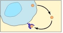
The contraction and expansion of the muscle.

http://cnx.org/content/m46635/latest/2114_Skeletal_Muscle_Vein_Pump.jpg

My 2nd hit is with Endothelial cells (ECs). These cells promote muscle relaxation through the release of prostaglandins, and endothelium-derived hyperpolarizing factors (EDHFs). These paracrine factors promote K+ efflux from muscle cells, with ensuing relaxation. Recent evidence suggests that ECs may also effect muscle relaxation via direct myoendothelial coupling. Regions of close apposition between endothelium and muscle cells are believed to contain myoendothelial gap junctions that promote the direct transfer of electrical and chemical signals between endothelial cells and muscle cells. Membrane potential has typically been measured in a single cell, with electrical coupling inferred between these respective cell types.

My communication with nerve cells.

Not only do I communicate with my fellow muscle cells but I can communicate with other cells through autocrine signalling, such as nerve cells. This occurs when a cell secretes signal molecules that can bind back to its own receptors. During my development, for example, once I am directed along a particular pathway of differentiation, I can secrete autocrine signals to myself that will reinforce this decision. 

Image

Cell producing a signaling molecule to which it responds.

Autocrine signaling is most effective when it is performed simultaneously by neighboring cells of the same type, and it is likely to be used to encourage groups of identical cells to make the same developmental decisions. As a result, the autocrine signaling mechanism is known as the “community effect”. Being that I am in close range to my fellow nerve cell, I am able to respond to a differentiation inducing signal which allows me to actively communicate with it, rather if I were alone and isolated. A group of us cells will produce a higher concentration of a secreted signal than if we were to work alone. When this signal binds back to a receptor on the same cell type, it encourages us, the cells to respond coordinately as a group.

Image

http://www.ncbi.nlm.nih.gov/books/NBK26813/figure/A2751/?report=objectonly

Autocrine signalling.

Cell to cell communication within the Xenopus Laevis.

Xenopus laevis

We cells have several different ways of communicating with each other, which depend on which type of cell we’re communicating with and also how far away that cell is. We can communicate by direct contact with each other, by using short range signals, by using long range signals or even by complex message in the form of electrical signals. All cells receive signals via signal receptor proteins, which may be inside the cell or on the cell membrane and they have a high affinity for binding with signal molecules. Once these signal molecules bind to their specific receptors, a signal cascade is usually set off and the cell reacts to this. Muscle cells, such as myself, are immediately surrounded by other muscle cells and together we make up muscle tissue. In the neighbouring areas, a variety of of types of cells exist, such as blood cells and nerve cells and I communicate with them very often.

As a muscle cell, I am surrounded by cells of the same type and we communicate with each other via direct cell-cell interactions using a mechanism called Gap Junctions. Gap junctions are specialized tube-like junctions, filled with water, that can form between cells in direct contact with each other, connecting the cytoplasm of the cells. Using these gap junctions, I can communicate with other muscle cells around me by sending, or receiving, small signal molecules and/or ions that help in determining the process I, or others, have to perform, whether it is growth or work.

As a mature muscle cell, I can send signals directly to neighbouring cells thereby causing an effect or change in the internal composition in relation to the signals I send. For example, as muscle cells in the leg of Xenopus laevis, my neighbours and I must be able to utilize energy stores quickly and efficiently for the muscle to perform its function, which relates to movement in the organism. Therefore, we have many mitochondria in us to quickly produce energy for us to work. The mitochondria that we contain are signalled when they are needed to perform their function, mainly by an increase in Ca2+ levels inside of our cytoplasm. Using gap junctions, Ca2+ can be transferred from cell to cell easily and quickly and so can signal the start of work in many cells in a relatively short time.

Skeletal muscle cell

Skeletal muscle cell

How did I originate?

In the first part of my journey, I form through the Primordial Oogonium. This then leads to the process of Oocytogenesis, in which my primary self (Oocyte) is formed. Meiosis I then occurs forming my first polar body and my secondary Oocyte self.

My purpose within the Xenopus laevis is to first divide meiotically. After my 2nd meiotic division, I will stop developing and completion of my future self (the ovum) will be paused (in Metaphase II called Dictyate) until I am to be fertilized. This process occurs as follows (highlighted):

Oogonium —> (Oocytogenesis) —> Primary Oocyte —> (Meiosis I) —> First Polar Body (Discarded afterward) + Secondary oocyte —>  (Meiosis II) —> Second Polar Body (Discarded afterward) + Ovum.

When I get older, my main goal is to become fertilized and ultimately grow into a fully functioning organism, such as the one I currently reside within.

I am an active site for RNA and protein synthesis. My structure comprises of:

  • Cytoplasm.

I am rich in cytoplasm which contains yolk granules to nourish myself, early in development.

  • A Nucleus.

During my stage of oogenesis, my nucleus is called a germinal vesicle, which stores my genetic material.

  • A Nest.

The space wherein I am located in my immature state is the cell-nest.

  • Zona pellucida.

The zona pellucida protects me during my development.

Structure of an Oocyte

References:

1) “A summary of oogenesis in Xenopus laevis.” Dept. of Biology, University of Utah. The University of Utah, n.d. Web. 22 Sept. 2013. http://biologylabs.utah.edu/gard/html/Oogenesis/Oogenesis_body.htm

2) http://www.dartmouth.edu/~anatomy/Histo/lab_6/female/DMS174/29.gif

3) http://php.med.unsw.edu.au/embryology/images/6/65/Ovary_histology_061.jpg

My Model Organism.

My model organism is the Xenopus laevis –The African clawed frog. The Xenopus is also commonly referred to as the African clawed toadAfrican claw-toed frog or the platanna. The genus Xenopus is the only frog with clawed toes. The African clawed frog has a flat body with a relatively small head.  Its skin is smooth, with dorsal surfaces usually colored in mottled hues of olive-brown or gray with darker marks and ventral surfaces a creamy white color.  This frog has no tongue, no teeth, no eyelids, and no external eardrums.  Its forelimbs have four unwebbed fingers and its hind limbs have five long, webbed toes with dark claws on the three outer toes.

A close up of the Xenopus laevis

A close up of the Xenopus laevis

The Xenopus laevis' forelimbs (left) and hind limbs (right)

The Xenopus laevis’ forelimbs (left) and hind limbs (right)

References:

1) Willigan, Erin. “Introduced Species Summary Project – Xenopus laevis.”Columbia University in the City of New York. N.p., 20 Oct. 2001. Web. 26 Sept. 2013. http://www.columbia.edu/itc/cerc/danoff-burg/invasion_bio/inv_spp_summ/xenopus_laevis.htm

2) http://www.critterzone.com/animal-pictures-nature/stock-photos/Head-face-closeup-African-clawed-frog-AWAM080508-47.jpg

3) http://www.critterzone.com/animal-pictures-nature/stock-photos/Claw-foot-African-clawed-frog-Xenopus-laevis-AWAM080508-12.jpg

4) http://wwwdelivery.superstock.com/WI/223/4201/PreviewComp/SuperStock_4201-23296.jpg

What is a Model Organism?

Perhaps?

A model organism can be defined as a species that has been widely studied, usually because it is easy to maintain and breed in a laboratory setting and has particular experimental advantages. Model organisms are used to obtain information about other species, including humans that are more difficult to study directly.

Why use a model organism?

Many of the things we study in Biology, such as disease, development and genetics needs to be studied in vivo, to see how pathways and signals, for example, really work. Realistically always studying these in humans would be extremely expensive as well as potentially unsafe and unethical. We use model organisms instead as they provide amazing insight that cannot be gained from lab equipment. What we learn about that animal can then be used to predict what happens in other animals. This is because all living organisms have evolved from the same ancestor and many pathways are the same across different species. In addition, model organisms are readily available, can be easily manipulated and display rapid development with their short life cycles.

Model organisms and their link to humans

Model organisms and their link to humans

References:

1) Twyman, Richard. “What are model organisms? | The Human Genome .” In the genome | The Human Genome . N.p., 28 Aug. 2002. Web. 21 Sept. 2013. http://genome.wellcome.ac.uk/doc_WTD020803.html

2)  “Using Model Organisms to Study Health and Disease – National Institute of General Medical Sciences.” NIGMS Home – National Institute of General Medical Sciences. N.p., 12 June 2013. Web. 21 Sept. 2013. http://www.nigms.nih.gov/Education/modelorg_factsheet.htm

3) http://www.prokop.co.uk/Research/LAYMAN/model-animals.gif

New beginnings -Cell and Developmental Biology.

Initially this blog began as an outlet for Biochemistry information. However, as my degree progresses, so too, my courses vary. The posts that follow this will entail an in-depth understanding of developmental anatomy. It will consequently detail the life of a specified cell present within a model organism.

Stay tuned for my model organism, in which I will reside!

Video Review #2-DNA and Nucleotides

Video link: Crashcourse 2012. “DNA Structure and Replication: Crash Course Biology #10.”  http://www.youtube.com/watch?v=8kK2zwjRV0M

Firstly, in the video, he speaks of DNA and what it encompasses in a brief point form manner. He states that nucleic acids are polymers, each one consists of many small repeating units. In DNA, they are referred to as Nucleotides; linked together they form polynucleotides. Next, he explains what a DNA molecule is made of (5-Carbon Sugar Molecule, Phosphate Group and 1 of 4 Nitrogen Bases). Of course you already knew this! The double helix is pair of molecules held tightly together, like a ladder. The sugar molecules & phosphates bind together thus forming the sugar-phosphate backbone at each side of helix, in opposite directions.

He continues to explain the concept of Directionality in a nucleic acid; the 5′-end and the 3′-end and vice-versa. This is explained in detail in the slides provided on MyElearning. The nitogenous bases can only be bonded to it’s specific partner, if you will. Adenine to Tryosine and Guanine to Cytosine, the base pairs. This concept is of great importantance in base sequencing. Next he speaks about RNA and how it differs to a DNA molecule. One such difference lies in its base pairs. Replication, as he speaks about next, is done by the “unzipping” of the double helix by the enzyme Helicase. You should be able to identify the “replication fork”, “leading strand” and the “lagging strand” on a basic diagram. These unwound sections are used as newly created templates, which will be used to create 2 complementary DNA strands leading in opposite directions.

DNA polymerase, adds matching nucleotides onto the main stem all the way down the molecule. The DNA polymerase is assisted in the beginning by RNA Primase, as it is only needed then. The DNA polymerase then follows suit and adds the rest of the bases to the leading strand. For the lagging strand, the process is slightly different. The DNA Polymerase can only copy strands in the 5′-3′ direction. The lagging strand however, is 3′-5′. DNA Polymerase therefore can only add new nucleotides to the 3 3′ end. RNA Primase, again enters at the beginning, the DNA Polymerase then enters moving backwards on the strand. DNA ligase join these fragments together, adhering the bonds to one another.

Throughtout the video he makes quirky remarks and comments as well as gives us fun and interesting facts through his “Biolo-graphy” session. Who knew that there were 6 Billion base pairs in EVERY cell! I certainly didn’t! Also, he gave a spontaneous pop quiz after the first 5 minutes of his lecture, a great way to maintain our focus! I was able to retain the information he explained, as I hope you will too!

Electron Transport Chain Video Review #1

Video link: Khan Academy. 2009. “Electron Transport Chain” http://www.youtube.com/watch?v=mfgCcFXUZRk

After viewing this video, I gathered that after glycolysis, 10 NADH and 2 FADHare left.  These are used in the Electron Transport Chain. This process is used to generate ATP. In the video, he states that the NADH is indirectly responsible for 3 ATP and each FADH2 is indirectly responsible for  the generation 2 ATP molecules. This is because the electrons that are entering the ETC are at a slightly lower energy level than the ones of NADH. The oxidation of NADH (NADH—->NAD+ + H+ + 2e) is the 1st step of the ETC. The last step involves (2e + H+ + ½ O—-> H2O) the reduction of oxygen to water; the oxidation of NADH to NAD+.

The 2e- used in oxidation then gets transported to a series of transition molecules, entering slightly lower energy states.  They are then used in the reduction of oxygen to water. When an electron goes from a higher energy state to a lower energy state, it releases energy.This energy is used to pump protons across the membrane of the cristae of the mitochondria. The oxidation and reduction processes occur in protein complexes located in the matrix of the mitochondria. When these proteins release energy, it is used to pump Hydrogen protons in particular into the outer membrane. The oxidation of NADH releases its by-product.  As a result, the outer membrane becomes more acidic than the matrix. An electric gradient/potential is the created between the outer (positive) membrane and the inner (negative) membrane.  

When this gradient forms, the hydrogen protons try to re-enter the matrix. ATP formation occurs in the cristae via the protein ATP synthase. These hydrogen ions enter the inner matrix via ATP synthase. This axle like structure on the top of the matrix as well as an extended part at the bottom allows the ions to enter via the spinning of the axle on the top. An ADP molecule and its 2 phosphate groups attach to 1 part  of the protein. The phosphate also attaches to another part of the protein. As the inner axle turns, the outer housing of the membrane due to electrical charges will squeeze the ADP and the phosphate together to form ATP. This occurs on 3 different sites simultaneously producing 3 ATP.

ATP Synthase

Summary:

Electrons are moving from the NADH and the FADH2 to essentially reduce O2. As they do this, they release energy as they go from 1 molecule to another. This energy is used to pump Hydrogen protons into the outer compartment of the mitochondria. The gradient created, makes the hydrogen protons want to enter the inner matrix. As they re-enter, this force drives the ATP Synthase “engine” which produces the ATP.

Left from Glycolysis:

10 NADH —-> 30 ATP

2 FADH2 —–> 4 ATP

Glycolysis and Krebs Cycle produces:

4 ATP molecules.

This amounts to 38 ATP molecules, from 1 molecule of Glucose.