Part III -Long distance-cell to cell communication in Xenopus Laevis.

For the last part of this blog, I will investigate long distance cell to cell communication between myself, a muscle cell and 2 other interrelated cells within myself.  Long distance signaling are cellular signals which are carried over long distances in the body called endocrine signaling. Endocrine signals employ hormones which are produced by endocrine cells. They travel through the blood to reach all parts of the body. Most hormones initiate a cellular response by initially combining with either a specific intracellular or cell membrane associated receptor protein. Specificity of signaling can be controlled if only some cells can respond to a particular hormone. A cell may have several different receptors that recognize the same hormone and activate different signal transduction pathways, or a cell may have several different receptors that recognize different hormones and activate the same biochemical pathway. For many hormones, including most protein hormones, the receptor is membrane-associated and embedded in the plasma membrane at the surface of the cell.

My 1st hit is with the cell of the hypothalamus. When an organism is threatened physically or emotionally, the hypothalamus readies the body to “fight” or “take flight” by sending impulses to the adrenal medulla. In response, the medulla secretes norepinephrine (in small amounts) and epinephrine (in larger amounts). Norepinephrine causes blood vessels in the skin and skeletal muscles to constrict, raising blood pressure. It is then that my hit is undertaken. Epinephrine causes an increase in heart rate and contraction, stimulates the liver to change glycogen to glucose for use as energy by the cells, and stimulates fatty tissue to break down and release stored fats for use as energy by the cells as well. The actions of both hormones bring about increased levels of oxygen and glucose in the blood and a faster circulation of blood to the body organs, especially the brain, muscles, and heart. Reflexes and body movements quicken and the body is better able to handle a short-term emergency situation.

The contraction and expansion of the muscle.

http://cnx.org/content/m46635/latest/2114_Skeletal_Muscle_Vein_Pump.jpg

My 2nd hit is with Endothelial cells (ECs). These cells promote muscle relaxation through the release of prostaglandins, and endothelium-derived hyperpolarizing factors (EDHFs). These paracrine factors promote K+ efflux from muscle cells, with ensuing relaxation. Recent evidence suggests that ECs may also effect muscle relaxation via direct myoendothelial coupling. Regions of close apposition between endothelium and muscle cells are believed to contain myoendothelial gap junctions that promote the direct transfer of electrical and chemical signals between endothelial cells and muscle cells. Membrane potential has typically been measured in a single cell, with electrical coupling inferred between these respective cell types.

How did I originate?

In the first part of my journey, I form through the Primordial Oogonium. This then leads to the process of Oocytogenesis, in which my primary self (Oocyte) is formed. Meiosis I then occurs forming my first polar body and my secondary Oocyte self.

My purpose within the Xenopus laevis is to first divide meiotically. After my 2nd meiotic division, I will stop developing and completion of my future self (the ovum) will be paused (in Metaphase II called Dictyate) until I am to be fertilized. This process occurs as follows (highlighted):

Oogonium —> (Oocytogenesis) —> Primary Oocyte —> (Meiosis I) —> First Polar Body (Discarded afterward) + Secondary oocyte —>  (Meiosis II) —> Second Polar Body (Discarded afterward) + Ovum.

When I get older, my main goal is to become fertilized and ultimately grow into a fully functioning organism, such as the one I currently reside within.

I am an active site for RNA and protein synthesis. My structure comprises of:

  • Cytoplasm.

I am rich in cytoplasm which contains yolk granules to nourish myself, early in development.

  • A Nucleus.

During my stage of oogenesis, my nucleus is called a germinal vesicle, which stores my genetic material.

  • A Nest.

The space wherein I am located in my immature state is the cell-nest.

  • Zona pellucida.

The zona pellucida protects me during my development.

Structure of an Oocyte

References:

1) “A summary of oogenesis in Xenopus laevis.” Dept. of Biology, University of Utah. The University of Utah, n.d. Web. 22 Sept. 2013. http://biologylabs.utah.edu/gard/html/Oogenesis/Oogenesis_body.htm

2) http://www.dartmouth.edu/~anatomy/Histo/lab_6/female/DMS174/29.gif

3) http://php.med.unsw.edu.au/embryology/images/6/65/Ovary_histology_061.jpg

Artificial jellyfish built from rat cells.

I came across this article while looking for a published paper. This unusual concept caught my attention, so I thought I would share it with everyone :).

Bioengineers have made an artificial jellyfish using silicone and muscle cells from a rat’s heart. The synthetic creature, dubbed a medusoid, looks like a flower with eight petals. When placed in an electric field, it pulses and swims exactly like its living counterpart.

“Morphologically, we’ve built a jellyfish. Functionally, we’ve built a jellyfish. Genetically, this thing is a rat,” says Kit Parker, a biophysicist at Harvard University in Cambridge, Massachusetts, who led the work. The project is described today in Nature Biotechnology1.

Parker’s lab works on creating artificial models of human heart tissues for regenerating organs and testing drugs, and the team built the medusoid as a way of understanding the “fundamental laws of muscular pumps”. It is an engineer’s approach to basic science: prove that you have identified the right principles by building something with them.


A jellyfish made of silicone and rat heart cells ‘swims’ in water when subjected to an electric field. In 2007, Parker was searching for new ways of studying muscular pumps when he visited the New England Aquarium in Boston, Massachusetts. “I saw the jellyfish display and it hit me like a thunderbolt,” he says. “I thought: I know I can build that.” To do so, he recruited John Dabiri, a bioengineer who studies biological propulsion at the California Institute of Technology (Caltech) in Pasadena. “I grabbed him and said, ‘John, I think I can build a jellyfish.’ He didn’t know who I was, but I was pretty excited and waving my arms, and I think he was afraid to say no.”

Janna Nawroth, a graduate student at Caltech who performed most of the experiments, began by mapping every cell in the bodies of juvenile moon jellies (Aurelia aurita) to understand how they swim. A moon jelly’s bell consists of a single layer of muscle, with fibres that are tightly aligned around a central ring and along eight spokes.

To make the bell beat downwards, electrical signals spread through the muscle in a smooth wave, “like when you drop a pebble in water”, says Parker. “It’s exactly like what you see in the heart. My bet is that to get a muscular pump, the electrical activity has got to spread as a wavefront.”

Form and function.

Nawroth created a structure with the same properties by growing a single layer of rat heart muscle on a patterned sheet of polydimethylsiloxane. When an electric field is applied across the structure, the muscle contracts rapidly, compressing the medusoid and mimicking a jellyfish’s power stroke. The elastic silicone then pulls the medusoid back to its original flat shape, ready for the next stroke.

When placed between two electrodes in water, the medusoid swam like the real thing. It even produced water currents similar to those that wash food particles into jellyfish’s mouths. “We thought if we’re really good at this, we’re going to recreate that vortex, and we did,” says Parker. “We took a rat apart and rebuilt it as a jellyfish.”

“I think that this is terrific,” says Joseph Vacanti, a tissue engineer at Massachusetts General Hospital in Boston. “It is a powerful demonstration of engineering chimaeric systems of living and non-living components.”

Parker says his team is taking synthetic biology to a new level. “Usually when we talk about synthetic life forms, somebody will take a living cell and put new genes in. We built an animal. It’s not just about genes, but about morphology and function.”

The team now plans to build a medusoid using human heart cells. The researchers have filed a patent to use their design, or something similar, as a platform for testing drugs. “You’ve got a heart drug?” says Parker. “You let me put it on my jellyfish, and I’ll tell you if it can improve the pumping.”

They also hope to reverse-engineer other marine life forms, says Parker. “We’ve got a whole tank of stuff in there, and an octopus on order.”

Quiz #1-The Cell

We were first tested on Wednesday 30th January on the Cell in groups of 5. The test was composed of 10 Multiple Choice questions which were fairly easy to answer. My group however received 9/10 even though we answered all of the questions correctly, to our knowledge. We still are not sure as to why we received this mark. Nevertheless, I enjoy partaking in these quizzes firstly because it’s easy marks to obtain and also because it allows me to keep on top of my work for I now expect a quiz on a weekly basis which encourages me to study even more!

Reflection on the cell.

A cell may seem simple to define. However, valuable information can be left out in the process, rendering the explanation insufficient and incomplete. It can be defined as “The smallest unit capable of performing life functions.” This definition is too basic. What are these life functions? They include growth, metabolism, response to stimuli and replication. With immense help from Mr.Matthew’s YouTube videos, I was able to understand and correctly describe the structure and function of many organelles such as the cytoskeleton, lysosomes, proteasomes and ribosomes to name a few.

During this revision, I realized that I had forgotten some of the organelles’ functions, specifically the Endoplasmic Reticulum, which happens to be one of my favorite words in the English language. I now recall that the ER is a network of membranous tubules within the cytoplasm of a eukaryotic cell, continuous with the nuclear membrane. It usually has ribosomes attached and is involved in protein and lipid synthesis via the rough endoplasmic reticulum.

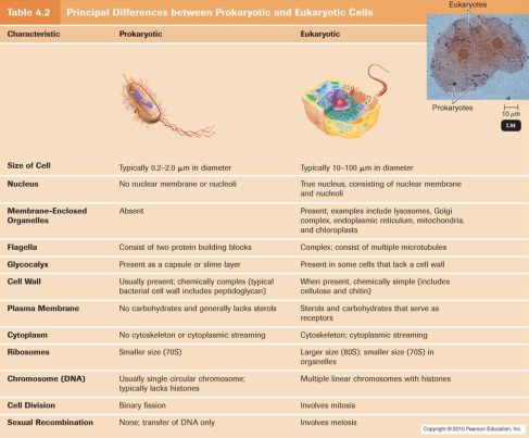
Mr. Matthew’s videos and lectures also encouraged me to do further research on sub-topics which I would not have on my own. One such example is with respect to Ribosomes. In prokaryotic cells, ribosomes are 70 s whereas in Eukaryotic cells, ribosomes are 80 s. But what does the “s” stand for? I have never questioned this before my lecture on Wednesday 23rd January, a fault of my own. Nevertheless, the “s” refers to svedberg unit (symbol S, sometimes Sv). Which is a non-SI unit for sedimentation rate. The sedimentation rate is the rate at which particles of a given size and shape travel to the bottom of the tube under centrifugal force.This reflects the rate at which a molecule sediments under the centrifugal force of a centrifuge.The svedberg is technically a measure of time, and is defined as exactly 10−13 seconds (100 fs)

[For some of you who did not heed Mr. Matthew’s gesture to research this]

In this lecture, we were also taught the differences between a Eukaryotic and a Prokaryotic cell, as well as the differences and similarities among a plant and animal cell. I have attached images below to illustrate these.

Image

Image