How did I originate?

In the first part of my journey, I form through the Primordial Oogonium. This then leads to the process of Oocytogenesis, in which my primary self (Oocyte) is formed. Meiosis I then occurs forming my first polar body and my secondary Oocyte self.

My purpose within the Xenopus laevis is to first divide meiotically. After my 2nd meiotic division, I will stop developing and completion of my future self (the ovum) will be paused (in Metaphase II called Dictyate) until I am to be fertilized. This process occurs as follows (highlighted):

Oogonium —> (Oocytogenesis) —> Primary Oocyte —> (Meiosis I) —> First Polar Body (Discarded afterward) + Secondary oocyte —>  (Meiosis II) —> Second Polar Body (Discarded afterward) + Ovum.

When I get older, my main goal is to become fertilized and ultimately grow into a fully functioning organism, such as the one I currently reside within.

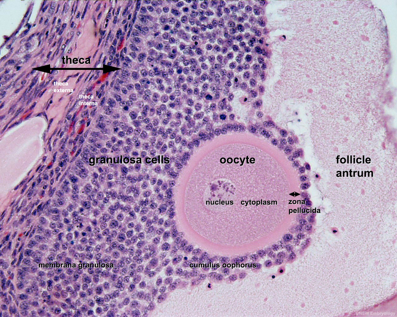
I am an active site for RNA and protein synthesis. My structure comprises of:

  • Cytoplasm.

I am rich in cytoplasm which contains yolk granules to nourish myself, early in development.

  • A Nucleus.

During my stage of oogenesis, my nucleus is called a germinal vesicle, which stores my genetic material.

  • A Nest.

The space wherein I am located in my immature state is the cell-nest.

  • Zona pellucida.

The zona pellucida protects me during my development.

Structure of an Oocyte

References:

1) “A summary of oogenesis in Xenopus laevis.” Dept. of Biology, University of Utah. The University of Utah, n.d. Web. 22 Sept. 2013. http://biologylabs.utah.edu/gard/html/Oogenesis/Oogenesis_body.htm

2) http://www.dartmouth.edu/~anatomy/Histo/lab_6/female/DMS174/29.gif

3) http://php.med.unsw.edu.au/embryology/images/6/65/Ovary_histology_061.jpg

My identity revealed.

This is my life! I am an oocyte or egg cell. I am a female gametocyte involved in sexual reproduction. I am contained within the ovarian follicle which is enclosed within the ovary.

A secondary oocyte

A secondary oocyte

Reference:

1) http://www.clipart.dk.co.uk/DKImages/exp_humanbody/exp_human002.jpg

My Model Organism.

My model organism is the Xenopus laevis –The African clawed frog. The Xenopus is also commonly referred to as the African clawed toadAfrican claw-toed frog or the platanna. The genus Xenopus is the only frog with clawed toes. The African clawed frog has a flat body with a relatively small head.  Its skin is smooth, with dorsal surfaces usually colored in mottled hues of olive-brown or gray with darker marks and ventral surfaces a creamy white color.  This frog has no tongue, no teeth, no eyelids, and no external eardrums.  Its forelimbs have four unwebbed fingers and its hind limbs have five long, webbed toes with dark claws on the three outer toes.

A close up of the Xenopus laevis

A close up of the Xenopus laevis

The Xenopus laevis' forelimbs (left) and hind limbs (right)

The Xenopus laevis’ forelimbs (left) and hind limbs (right)

References:

1) Willigan, Erin. “Introduced Species Summary Project – Xenopus laevis.”Columbia University in the City of New York. N.p., 20 Oct. 2001. Web. 26 Sept. 2013. http://www.columbia.edu/itc/cerc/danoff-burg/invasion_bio/inv_spp_summ/xenopus_laevis.htm

2) http://www.critterzone.com/animal-pictures-nature/stock-photos/Head-face-closeup-African-clawed-frog-AWAM080508-47.jpg

3) http://www.critterzone.com/animal-pictures-nature/stock-photos/Claw-foot-African-clawed-frog-Xenopus-laevis-AWAM080508-12.jpg

4) http://wwwdelivery.superstock.com/WI/223/4201/PreviewComp/SuperStock_4201-23296.jpg

What is a Model Organism?

Perhaps?

A model organism can be defined as a species that has been widely studied, usually because it is easy to maintain and breed in a laboratory setting and has particular experimental advantages. Model organisms are used to obtain information about other species, including humans that are more difficult to study directly.

Why use a model organism?

Many of the things we study in Biology, such as disease, development and genetics needs to be studied in vivo, to see how pathways and signals, for example, really work. Realistically always studying these in humans would be extremely expensive as well as potentially unsafe and unethical. We use model organisms instead as they provide amazing insight that cannot be gained from lab equipment. What we learn about that animal can then be used to predict what happens in other animals. This is because all living organisms have evolved from the same ancestor and many pathways are the same across different species. In addition, model organisms are readily available, can be easily manipulated and display rapid development with their short life cycles.

Model organisms and their link to humans

Model organisms and their link to humans

References:

1) Twyman, Richard. “What are model organisms? | The Human Genome .” In the genome | The Human Genome . N.p., 28 Aug. 2002. Web. 21 Sept. 2013. http://genome.wellcome.ac.uk/doc_WTD020803.html

2)  “Using Model Organisms to Study Health and Disease – National Institute of General Medical Sciences.” NIGMS Home – National Institute of General Medical Sciences. N.p., 12 June 2013. Web. 21 Sept. 2013. http://www.nigms.nih.gov/Education/modelorg_factsheet.htm

3) http://www.prokop.co.uk/Research/LAYMAN/model-animals.gif

New beginnings -Cell and Developmental Biology.

Initially this blog began as an outlet for Biochemistry information. However, as my degree progresses, so too, my courses vary. The posts that follow this will entail an in-depth understanding of developmental anatomy. It will consequently detail the life of a specified cell present within a model organism.

Stay tuned for my model organism, in which I will reside!