Glycolysis breakdown.

What is Glycolysis?

Glycolysis means the splitting of sugar. In glycolysis, glucose (a 6 carbon sugar) is split into 2 molecules of a 3-carbon sugar. Glycolysis yields 2 molecules of ATP (free energy containing molecule), 2 molecules of pyruvic acid & 2 “high energy” electron carrying molecules of NADH. Glycolysis can occur with (aerobically) or without oxygen (anaerobically). In the presence of oxygen, glycolysis is the first stage of cellular respiration. Without oxygen, glycolysis allows cells to make small amounts of ATP. This process is called fermentation.

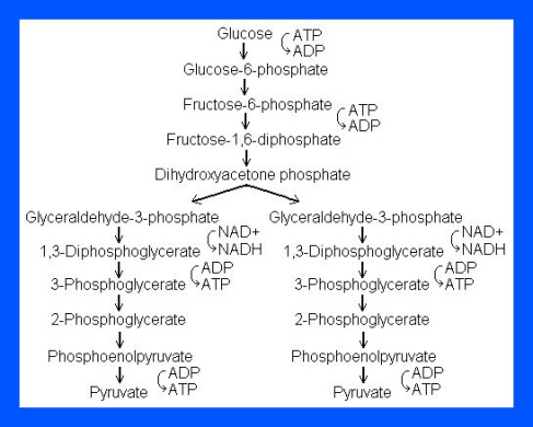
The process of Glycolysis occurs through 10 steps.

Step 1

The enzyme hexokinase phosphorylates (adds a phosphate group to) glucose in the cell’s cytoplasm. In the process, a phosphate group from ATP is transferred to glucose producing glucose 6-phosphate.

  • Glucose (C6H12O6) + hexokinase + ATP → ADP + Glucose 6-phosphate (C6H11O6P1

Step 2

The enzyme phosphohexose isomerase converts glucose 6-phosphate into its isomer fructose 6-phosphate. Isomers have the same molecular formula, but the atoms of each molecule are arranged differently.

  • Glucose 6-phosphate (C6H11O6P1) + Phosphoglucoisomerase → Fructose 6-phosphate (C6H11O6P1)

Step 3

The enzyme phosphofructokinase-1 uses another ATP molecule to transfer a phosphate group to fructose 6-phosphate to form fructose 1, 6-bisphosphate.

  • Fructose 6-phosphate (C6H11O6P1) + phosphofructokinase + ATP → ADP + Fructose 1, 6-bisphosphate (C6H10O6P2

Step 4

The enzyme aldolase splits fructose 1, 6-bisphosphate into two sugars that are isomers of each other. These two sugars are dihydroxyacetone phosphate and glyceraldehyde phosphate.

  • Fructose 1, 6-bisphosphate (C6H10O6P2) + aldolase → Dihydroxyacetone phosphate (C3H5O3P1) + Glyceraldehyde phosphate (C3H5O3P1)

Step 5

The enzyme triose phosphate isomerase rapidly inter-converts the molecules dihydroxyacetone phosphate and glyceraldehyde phosphate. Glyceraldehyde phosphate is removed as soon as it is formed to be used in the next step of glycolysis.

  • Dihydroxyacetone phosphate (C3H5O3P1) → Glyceraldehyde phosphate (C3H5O3P1

Net result for steps 4 and 5: Fructose 1, 6-bisphosphate (C6H10O6P2) ↔ 2 molecules of Glyceraldehyde phosphate (C3H5O3P1)

Step 6

The enzyme Glyceraldehyde 3-phosphate dehydrogenase serves two functions in this step. First the enzyme transfers a hydrogen (H) from glyceraldehyde phosphate to the oxidizing agent nicotinamide adenine dinucleotide (NAD+) to form NADH. Next triose phosphate dehydrogenase adds a phosphate (P) from the cytosol to the oxidized glyceraldehyde phosphate to form 1, 3-bisphosphoglycerate. This occurs for both molecules of glyceraldehyde phosphate produced in Step 5.

  • Triose phosphate dehydrogenase + 2 H + 2 NAD+ → 2 NADH + 2 H+
  • Triose phosphate dehydrogenase + 2 P + 2 glyceraldehyde phosphate (C3H5O3P1) → 2 molecules of 1,3-bisphosphoglycerate (C3H4O4P2

Step 7

The enzyme phosphoglycerate kinase transfers a P from 1,3-bisphosphoglycerate to a molecule of ADP to form ATP. This happens for each molecule of 1,3-bisphosphoglycerate. The process yields two 3-phosphoglycerate molecules and two ATP molecules.

  • 2 molecules of 1,3-bisphoshoglycerate (C3H4O4P2) + phosphoglycerate kinase + 2 ADP → 2 molecules of 3-phosphoglycerate (C3H5O4P1) + 2 ATP 

Step 8

The enzyme phosphoglycerate mutase relocates the P from 3-phosphoglycerate from the third carbon to the second carbon to form 2-phosphoglycerate.

  • 2 molecules of 3-Phosphoglycerate (C3H5O4P1) + phosphoglyceromutase → 2 molecules of 2-Phosphoglycerate (C3H5O4P1)

Step 9

The enzyme enolase removes a molecule of water from 2-phosphoglycerate to form phosphoenolpyruvic acid (PEP). This happens for each molecule of 2-phosphoglycerate.

  • 2 molecules of 2-Phosphoglycerate (C3H5O4P1) + enolase → 2 molecules of phosphoenolpyruvic acid (PEP) (C3H3O3P1

Step 10

The enzyme pyruvate kinase transfers a P from PEP to ADP to form pyruvic acid and ATP. This happens for each molecule of PEP. This reaction yields 2 molecules of pyruvic acid and 2 ATP molecules.

  • 2 molecules of PEP (C3H3O3P1) + pyruvate kinase + 2 ADP → 2 molecules of pyruvic acid (C3H4O3) + 2 ATP

In summary, a single glucose molecule in glycolysis produces a total of 2 molecules of pyruvic acid, 2 molecules of ATP, 2 molecules of NADH and 2 molecules of water. 

Although 2 ATP molecules are used in steps 1-3, 2 ATP molecules are generated in step 7 and 2 more in step 10. This gives a total of 4 ATP molecules produced. If you subtract the 2 ATP molecules used in steps 1-3 from the 4 generated at the end of step 10, you end up with a net total of 2 ATP molecules produced.

Image

Enzyme Specificity and Reversible Inhibition

Enzymes are biological catalysts that speed up a chemical reaction by providing an alternative pathway with a lower activation energy. One enzymatic property is that they are specific relative to the reactions they catalyze. Specificity can be classed into four types. Firstly, Absolute specificity occurs when the enzyme will catalyze a single reaction. Group specificity takes place when the enzyme will act only on molecules that have fixed functional groups, such as phosphate, amino and methyl groups. Linkage specificity is induced when the enzyme will act on a certain type of chemical bond disregarding of the rest of the structure. Lastly, Stereochemical specificity is when the enzyme will react on a specific steric or optical isomer.

Enzymes do not always follow a smooth pattern of operation. Enzyme inhibitors are molecules that interact with the enzyme to prevent it from working in its usual order. The types of inhibitors include: nonspecific, irreversible, reversible.

Reversible inhibition is categorized according to the effect of varying the concentration of the enzyme’s substrate on the inhibitor.

  • In competitive inhibition, the inhibitor and the substrate resemble each other, and therefore cannot bind to the enzyme at the same time. The inhibitor binds to the active site of the enzyme, thus slowing the rate of reaction. The apparent Km will increase because it takes a higher concentration of the substrate to reach the Km point or half the Vmax. A competitive inhibitor will increase the apparent KM value for its substrate with no change in the apparent Vmax value.
  • In uncompetitive inhibition, the inhibitor does not resemble the substrate in this type of inhibition. The inhibitor binds only to the substrate-enzyme complex. This type of inhibition causes Km and Vmax to decrease.
  • In mixed inhibition, the inhibitor can bind to the enzyme at similar times as the enzyme’s substrate. The binding of the substrate and vice versa is affected by the binding of the inhibitor. This type of inhibition generally results from an allosteric effect where the inhibitor binds to a different site on an enzyme. In this type of inhibition, Vmax decreases, while Km may increase or decrease.
  • Non-competitive inhibition is a form of mixed inhibition where the  but does not affect the binding of substrate. The inhibitor does not resemble the substrate. Vmax will decrease due to the inability for the reaction to proceed as efficiently, but Km will remain the same as the actual binding of the substrate, by definition, will still function properly.

Non-competitive Inhibitor

Lineweaver–Burk plots displaying the 4 types of reversible inhibition.

Enzyme Inhibition Equations

Enzyme Inhibition Equations

Published Paper #2

Article reference: Shen, Helen. 2012. “Enzymes grow artificial DNA” Accessed March 31, 2013. http://www.nature.com/news/enzymes-grow-artificial-dna-1.10487#/b1

Image

DNA, commonly known as the blueprint of life, is the naturally occurring hereditary material in humans and almost all organisms; or so we thought. Scientists have developed lab made alternatives of DNA. These variants act much like DNA in which these mechanisms allow it to store and transfer hereditary information. Then again why should these synthetic strands be crafted? According to Philipp Holliger, a synthetic biologist at the Medical Research Council Laboratory of Molecular Biology in Cambridge, UK, he states that these lab assembled DNA backbones can aid in the development of new drugs and nanotechnologies.

The proposition.

The construction of DNA composes of nucleic acid bases- A, C, G and T assembled on a phosphate and sugar deoxyribose backbone. These replicated polymers known as XNA’s carry the same lettered sequencing except for its backbone which is made of different sugars from the novel DNA molecules. Another biochemist, Gerald Joyce explained that while this task has been attempted before, they were unable to make additional copies these already generated XNA strands. However, recent developments have supplied a way, and a considerable one at that, says Eric Kool, a chemist at Stanford University in California. As a result, the successive DNA to XNA transmission have permitted researches to select the XNA’s that are attached to choice target proteins from a vast number of samples.

Innovation.

With the progression of such research, similar undertakings were undergone. Steven Banner, a biochemist at the Foundation for Applied Molecular Evolution in Florida, together with his associates have simulated polymers with additional artificial genetic lettering on an original DNA backbone. This synthetic sequencing can merge with the modified XNA backbone creating a structure resistant to chemical degradation.

The next step.

Upon further exploration, Biotechnologists may have discovered tools such as polymers that can bind and inhibit proteins, in these evolvable XNA’s. One such example includes strains involved in macular degeneration, a disease which affects older adults resulting in loss of vision in the center of the macula due to retinal damage.

Historical ingenuity put to use.

DNA and RNA, as simple as they may seem to be comprised of, are difficult to produce. Many researchers believe that another simpler molecule was derived first. The TNA3 molecule was developed by Albert Eschenmoser in the year 2000. This strand was in essence a XNA together with a α-l-threofuranosyl nucleic acid backbone and 1 of the 6 polymers used in the study.  This experiment exhibited that the TNAcould conform to the DNA by twisting into a double-helix spiral. Ramanarayanan Krishnamurthy, a leading synthetic organic chemist, noted that the enzymes produced by Holliger advanced past research “by leaps and bounds”. He stated that these developed enzymes confirm that DNA can indeed trade information efficiently with TNA and additional polymers apart from RNA.

Although the XNA’s are dependent on DNA derived enzymes to replicate, researchers can presently duplicate artificial genes resistant to biodegradation.

Additional reading:

References: