Video Review #2-DNA and Nucleotides

Video link: Crashcourse 2012. “DNA Structure and Replication: Crash Course Biology #10.”  http://www.youtube.com/watch?v=8kK2zwjRV0M

Firstly, in the video, he speaks of DNA and what it encompasses in a brief point form manner. He states that nucleic acids are polymers, each one consists of many small repeating units. In DNA, they are referred to as Nucleotides; linked together they form polynucleotides. Next, he explains what a DNA molecule is made of (5-Carbon Sugar Molecule, Phosphate Group and 1 of 4 Nitrogen Bases). Of course you already knew this! The double helix is pair of molecules held tightly together, like a ladder. The sugar molecules & phosphates bind together thus forming the sugar-phosphate backbone at each side of helix, in opposite directions.

He continues to explain the concept of Directionality in a nucleic acid; the 5′-end and the 3′-end and vice-versa. This is explained in detail in the slides provided on MyElearning. The nitogenous bases can only be bonded to it’s specific partner, if you will. Adenine to Tryosine and Guanine to Cytosine, the base pairs. This concept is of great importantance in base sequencing. Next he speaks about RNA and how it differs to a DNA molecule. One such difference lies in its base pairs. Replication, as he speaks about next, is done by the “unzipping” of the double helix by the enzyme Helicase. You should be able to identify the “replication fork”, “leading strand” and the “lagging strand” on a basic diagram. These unwound sections are used as newly created templates, which will be used to create 2 complementary DNA strands leading in opposite directions.

DNA polymerase, adds matching nucleotides onto the main stem all the way down the molecule. The DNA polymerase is assisted in the beginning by RNA Primase, as it is only needed then. The DNA polymerase then follows suit and adds the rest of the bases to the leading strand. For the lagging strand, the process is slightly different. The DNA Polymerase can only copy strands in the 5′-3′ direction. The lagging strand however, is 3′-5′. DNA Polymerase therefore can only add new nucleotides to the 3 3′ end. RNA Primase, again enters at the beginning, the DNA Polymerase then enters moving backwards on the strand. DNA ligase join these fragments together, adhering the bonds to one another.

Throughtout the video he makes quirky remarks and comments as well as gives us fun and interesting facts through his “Biolo-graphy” session. Who knew that there were 6 Billion base pairs in EVERY cell! I certainly didn’t! Also, he gave a spontaneous pop quiz after the first 5 minutes of his lecture, a great way to maintain our focus! I was able to retain the information he explained, as I hope you will too!

Electron Transport Chain Video Review #1

Video link: Khan Academy. 2009. “Electron Transport Chain” http://www.youtube.com/watch?v=mfgCcFXUZRk

After viewing this video, I gathered that after glycolysis, 10 NADH and 2 FADHare left.  These are used in the Electron Transport Chain. This process is used to generate ATP. In the video, he states that the NADH is indirectly responsible for 3 ATP and each FADH2 is indirectly responsible for  the generation 2 ATP molecules. This is because the electrons that are entering the ETC are at a slightly lower energy level than the ones of NADH. The oxidation of NADH (NADH—->NAD+ + H+ + 2e) is the 1st step of the ETC. The last step involves (2e + H+ + ½ O—-> H2O) the reduction of oxygen to water; the oxidation of NADH to NAD+.

The 2e- used in oxidation then gets transported to a series of transition molecules, entering slightly lower energy states.  They are then used in the reduction of oxygen to water. When an electron goes from a higher energy state to a lower energy state, it releases energy.This energy is used to pump protons across the membrane of the cristae of the mitochondria. The oxidation and reduction processes occur in protein complexes located in the matrix of the mitochondria. When these proteins release energy, it is used to pump Hydrogen protons in particular into the outer membrane. The oxidation of NADH releases its by-product.  As a result, the outer membrane becomes more acidic than the matrix. An electric gradient/potential is the created between the outer (positive) membrane and the inner (negative) membrane.  

When this gradient forms, the hydrogen protons try to re-enter the matrix. ATP formation occurs in the cristae via the protein ATP synthase. These hydrogen ions enter the inner matrix via ATP synthase. This axle like structure on the top of the matrix as well as an extended part at the bottom allows the ions to enter via the spinning of the axle on the top. An ADP molecule and its 2 phosphate groups attach to 1 part  of the protein. The phosphate also attaches to another part of the protein. As the inner axle turns, the outer housing of the membrane due to electrical charges will squeeze the ADP and the phosphate together to form ATP. This occurs on 3 different sites simultaneously producing 3 ATP.

ATP Synthase

Summary:

Electrons are moving from the NADH and the FADH2 to essentially reduce O2. As they do this, they release energy as they go from 1 molecule to another. This energy is used to pump Hydrogen protons into the outer compartment of the mitochondria. The gradient created, makes the hydrogen protons want to enter the inner matrix. As they re-enter, this force drives the ATP Synthase “engine” which produces the ATP.

Left from Glycolysis:

10 NADH —-> 30 ATP

2 FADH2 —–> 4 ATP

Glycolysis and Krebs Cycle produces:

4 ATP molecules.

This amounts to 38 ATP molecules, from 1 molecule of Glucose.

Citric Acid Cycle made simple

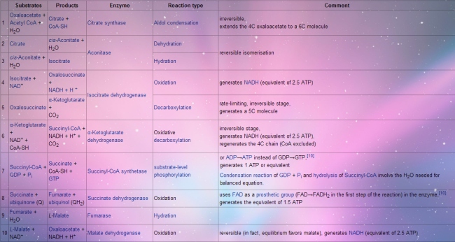
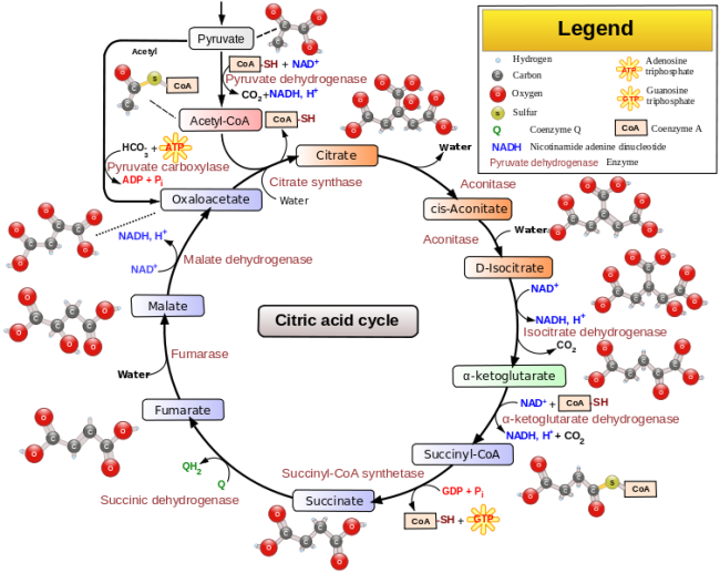
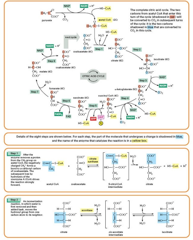
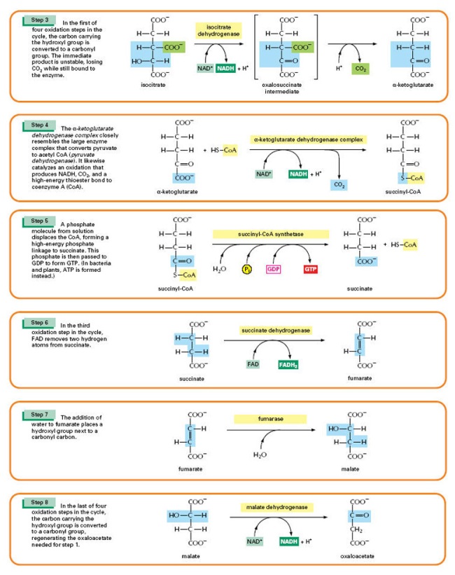
The citric acid cycle, also known as the Tricarboxylic acid cycle (TCA cycle), the Krebs cycle, or the Szent-Györgyi–Krebs cycle is a series of chemical reactions used by all aerobic organisms to generate energy through the oxidization of acetate derived from carbohydrates, fats and proteins into carbon dioxide. 

The following is an account of the Krebs cycle’s substrates, products, enzymes used and reaction type.

Image

Here are some additional diagrams and flow charts to better illustrate the mechanisms involved in the TCA Cycle.

Image

Image

Image

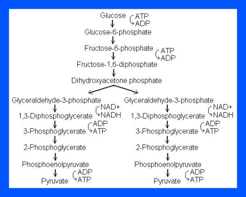
Glycolysis breakdown.

What is Glycolysis?

Glycolysis means the splitting of sugar. In glycolysis, glucose (a 6 carbon sugar) is split into 2 molecules of a 3-carbon sugar. Glycolysis yields 2 molecules of ATP (free energy containing molecule), 2 molecules of pyruvic acid & 2 “high energy” electron carrying molecules of NADH. Glycolysis can occur with (aerobically) or without oxygen (anaerobically). In the presence of oxygen, glycolysis is the first stage of cellular respiration. Without oxygen, glycolysis allows cells to make small amounts of ATP. This process is called fermentation.

The process of Glycolysis occurs through 10 steps.

Step 1

The enzyme hexokinase phosphorylates (adds a phosphate group to) glucose in the cell’s cytoplasm. In the process, a phosphate group from ATP is transferred to glucose producing glucose 6-phosphate.

  • Glucose (C6H12O6) + hexokinase + ATP → ADP + Glucose 6-phosphate (C6H11O6P1

Step 2

The enzyme phosphohexose isomerase converts glucose 6-phosphate into its isomer fructose 6-phosphate. Isomers have the same molecular formula, but the atoms of each molecule are arranged differently.

  • Glucose 6-phosphate (C6H11O6P1) + Phosphoglucoisomerase → Fructose 6-phosphate (C6H11O6P1)

Step 3

The enzyme phosphofructokinase-1 uses another ATP molecule to transfer a phosphate group to fructose 6-phosphate to form fructose 1, 6-bisphosphate.

  • Fructose 6-phosphate (C6H11O6P1) + phosphofructokinase + ATP → ADP + Fructose 1, 6-bisphosphate (C6H10O6P2

Step 4

The enzyme aldolase splits fructose 1, 6-bisphosphate into two sugars that are isomers of each other. These two sugars are dihydroxyacetone phosphate and glyceraldehyde phosphate.

  • Fructose 1, 6-bisphosphate (C6H10O6P2) + aldolase → Dihydroxyacetone phosphate (C3H5O3P1) + Glyceraldehyde phosphate (C3H5O3P1)

Step 5

The enzyme triose phosphate isomerase rapidly inter-converts the molecules dihydroxyacetone phosphate and glyceraldehyde phosphate. Glyceraldehyde phosphate is removed as soon as it is formed to be used in the next step of glycolysis.

  • Dihydroxyacetone phosphate (C3H5O3P1) → Glyceraldehyde phosphate (C3H5O3P1

Net result for steps 4 and 5: Fructose 1, 6-bisphosphate (C6H10O6P2) ↔ 2 molecules of Glyceraldehyde phosphate (C3H5O3P1)

Step 6

The enzyme Glyceraldehyde 3-phosphate dehydrogenase serves two functions in this step. First the enzyme transfers a hydrogen (H) from glyceraldehyde phosphate to the oxidizing agent nicotinamide adenine dinucleotide (NAD+) to form NADH. Next triose phosphate dehydrogenase adds a phosphate (P) from the cytosol to the oxidized glyceraldehyde phosphate to form 1, 3-bisphosphoglycerate. This occurs for both molecules of glyceraldehyde phosphate produced in Step 5.

  • Triose phosphate dehydrogenase + 2 H + 2 NAD+ → 2 NADH + 2 H+
  • Triose phosphate dehydrogenase + 2 P + 2 glyceraldehyde phosphate (C3H5O3P1) → 2 molecules of 1,3-bisphosphoglycerate (C3H4O4P2

Step 7

The enzyme phosphoglycerate kinase transfers a P from 1,3-bisphosphoglycerate to a molecule of ADP to form ATP. This happens for each molecule of 1,3-bisphosphoglycerate. The process yields two 3-phosphoglycerate molecules and two ATP molecules.

  • 2 molecules of 1,3-bisphoshoglycerate (C3H4O4P2) + phosphoglycerate kinase + 2 ADP → 2 molecules of 3-phosphoglycerate (C3H5O4P1) + 2 ATP 

Step 8

The enzyme phosphoglycerate mutase relocates the P from 3-phosphoglycerate from the third carbon to the second carbon to form 2-phosphoglycerate.

  • 2 molecules of 3-Phosphoglycerate (C3H5O4P1) + phosphoglyceromutase → 2 molecules of 2-Phosphoglycerate (C3H5O4P1)

Step 9

The enzyme enolase removes a molecule of water from 2-phosphoglycerate to form phosphoenolpyruvic acid (PEP). This happens for each molecule of 2-phosphoglycerate.

  • 2 molecules of 2-Phosphoglycerate (C3H5O4P1) + enolase → 2 molecules of phosphoenolpyruvic acid (PEP) (C3H3O3P1

Step 10

The enzyme pyruvate kinase transfers a P from PEP to ADP to form pyruvic acid and ATP. This happens for each molecule of PEP. This reaction yields 2 molecules of pyruvic acid and 2 ATP molecules.

  • 2 molecules of PEP (C3H3O3P1) + pyruvate kinase + 2 ADP → 2 molecules of pyruvic acid (C3H4O3) + 2 ATP

In summary, a single glucose molecule in glycolysis produces a total of 2 molecules of pyruvic acid, 2 molecules of ATP, 2 molecules of NADH and 2 molecules of water. 

Although 2 ATP molecules are used in steps 1-3, 2 ATP molecules are generated in step 7 and 2 more in step 10. This gives a total of 4 ATP molecules produced. If you subtract the 2 ATP molecules used in steps 1-3 from the 4 generated at the end of step 10, you end up with a net total of 2 ATP molecules produced.

Image

Multiple choice questions on Glycolysis.

Select the correct multiple answer by using ONE of the keys A, B, C, D or E below.

A) 1,2,3 are correct

B) 1 & 3 are correct

C) Only 4 is correct

D) 1 & 2 are correct

E) All are correct

1) Glycolytic pathway regulation involves:

1) Mg2+ as a cofactor.

2) Feedback, Product, Inhibition by ATP.

3) Allosteric simulation by ADP.

4) Allosteric inhibition by ATP.

5) All of the above.

2) Which of these enzymes regulates the glycolytic steps?

1)Phosphofructokinase.

2) Hexkinase.

3) Pyruvate Kinase.

4) Lactase.

3)Fructose-2,6 Bisphosphate:

1) Activates Phosphofructokinase.

2) Activates Glyceraldehyde-3-Phosphate.

3) Inhibits Fructose-1,6 Bisphosphate.

4) Inhibits Fructose-6-Phosphate.

4) Glucose from the breakdown of Glycogen is acquired from:

1) The muscles by phosphorolysis.

2) The liver by phosphorolysis.

3) The muscles by hydrolysis.

4) Feeder pathways.

5) Glycogen has:

1) α-1,4 linkages.

2) α-1,6 linkages.

3) α-1,4 and β-1,6 linkages.

4) α-1,4 and α-1,6 linkages.

5) α-1,6 and β-1,6 linkages.

Artificial jellyfish built from rat cells.

I came across this article while looking for a published paper. This unusual concept caught my attention, so I thought I would share it with everyone :).

Bioengineers have made an artificial jellyfish using silicone and muscle cells from a rat’s heart. The synthetic creature, dubbed a medusoid, looks like a flower with eight petals. When placed in an electric field, it pulses and swims exactly like its living counterpart.

“Morphologically, we’ve built a jellyfish. Functionally, we’ve built a jellyfish. Genetically, this thing is a rat,” says Kit Parker, a biophysicist at Harvard University in Cambridge, Massachusetts, who led the work. The project is described today in Nature Biotechnology1.

Parker’s lab works on creating artificial models of human heart tissues for regenerating organs and testing drugs, and the team built the medusoid as a way of understanding the “fundamental laws of muscular pumps”. It is an engineer’s approach to basic science: prove that you have identified the right principles by building something with them.


A jellyfish made of silicone and rat heart cells ‘swims’ in water when subjected to an electric field. In 2007, Parker was searching for new ways of studying muscular pumps when he visited the New England Aquarium in Boston, Massachusetts. “I saw the jellyfish display and it hit me like a thunderbolt,” he says. “I thought: I know I can build that.” To do so, he recruited John Dabiri, a bioengineer who studies biological propulsion at the California Institute of Technology (Caltech) in Pasadena. “I grabbed him and said, ‘John, I think I can build a jellyfish.’ He didn’t know who I was, but I was pretty excited and waving my arms, and I think he was afraid to say no.”

Janna Nawroth, a graduate student at Caltech who performed most of the experiments, began by mapping every cell in the bodies of juvenile moon jellies (Aurelia aurita) to understand how they swim. A moon jelly’s bell consists of a single layer of muscle, with fibres that are tightly aligned around a central ring and along eight spokes.

To make the bell beat downwards, electrical signals spread through the muscle in a smooth wave, “like when you drop a pebble in water”, says Parker. “It’s exactly like what you see in the heart. My bet is that to get a muscular pump, the electrical activity has got to spread as a wavefront.”

Form and function.

Nawroth created a structure with the same properties by growing a single layer of rat heart muscle on a patterned sheet of polydimethylsiloxane. When an electric field is applied across the structure, the muscle contracts rapidly, compressing the medusoid and mimicking a jellyfish’s power stroke. The elastic silicone then pulls the medusoid back to its original flat shape, ready for the next stroke.

When placed between two electrodes in water, the medusoid swam like the real thing. It even produced water currents similar to those that wash food particles into jellyfish’s mouths. “We thought if we’re really good at this, we’re going to recreate that vortex, and we did,” says Parker. “We took a rat apart and rebuilt it as a jellyfish.”

“I think that this is terrific,” says Joseph Vacanti, a tissue engineer at Massachusetts General Hospital in Boston. “It is a powerful demonstration of engineering chimaeric systems of living and non-living components.”

Parker says his team is taking synthetic biology to a new level. “Usually when we talk about synthetic life forms, somebody will take a living cell and put new genes in. We built an animal. It’s not just about genes, but about morphology and function.”

The team now plans to build a medusoid using human heart cells. The researchers have filed a patent to use their design, or something similar, as a platform for testing drugs. “You’ve got a heart drug?” says Parker. “You let me put it on my jellyfish, and I’ll tell you if it can improve the pumping.”

They also hope to reverse-engineer other marine life forms, says Parker. “We’ve got a whole tank of stuff in there, and an octopus on order.”

Enzyme Specificity and Reversible Inhibition

Enzymes are biological catalysts that speed up a chemical reaction by providing an alternative pathway with a lower activation energy. One enzymatic property is that they are specific relative to the reactions they catalyze. Specificity can be classed into four types. Firstly, Absolute specificity occurs when the enzyme will catalyze a single reaction. Group specificity takes place when the enzyme will act only on molecules that have fixed functional groups, such as phosphate, amino and methyl groups. Linkage specificity is induced when the enzyme will act on a certain type of chemical bond disregarding of the rest of the structure. Lastly, Stereochemical specificity is when the enzyme will react on a specific steric or optical isomer.

Enzymes do not always follow a smooth pattern of operation. Enzyme inhibitors are molecules that interact with the enzyme to prevent it from working in its usual order. The types of inhibitors include: nonspecific, irreversible, reversible.

Reversible inhibition is categorized according to the effect of varying the concentration of the enzyme’s substrate on the inhibitor.

  • In competitive inhibition, the inhibitor and the substrate resemble each other, and therefore cannot bind to the enzyme at the same time. The inhibitor binds to the active site of the enzyme, thus slowing the rate of reaction. The apparent Km will increase because it takes a higher concentration of the substrate to reach the Km point or half the Vmax. A competitive inhibitor will increase the apparent KM value for its substrate with no change in the apparent Vmax value.
  • In uncompetitive inhibition, the inhibitor does not resemble the substrate in this type of inhibition. The inhibitor binds only to the substrate-enzyme complex. This type of inhibition causes Km and Vmax to decrease.
  • In mixed inhibition, the inhibitor can bind to the enzyme at similar times as the enzyme’s substrate. The binding of the substrate and vice versa is affected by the binding of the inhibitor. This type of inhibition generally results from an allosteric effect where the inhibitor binds to a different site on an enzyme. In this type of inhibition, Vmax decreases, while Km may increase or decrease.
  • Non-competitive inhibition is a form of mixed inhibition where the  but does not affect the binding of substrate. The inhibitor does not resemble the substrate. Vmax will decrease due to the inability for the reaction to proceed as efficiently, but Km will remain the same as the actual binding of the substrate, by definition, will still function properly.

Non-competitive Inhibitor

Lineweaver–Burk plots displaying the 4 types of reversible inhibition.

Enzyme Inhibition Equations

Enzyme Inhibition Equations

Published Paper #2

Article reference: Shen, Helen. 2012. “Enzymes grow artificial DNA” Accessed March 31, 2013. http://www.nature.com/news/enzymes-grow-artificial-dna-1.10487#/b1

Image

DNA, commonly known as the blueprint of life, is the naturally occurring hereditary material in humans and almost all organisms; or so we thought. Scientists have developed lab made alternatives of DNA. These variants act much like DNA in which these mechanisms allow it to store and transfer hereditary information. Then again why should these synthetic strands be crafted? According to Philipp Holliger, a synthetic biologist at the Medical Research Council Laboratory of Molecular Biology in Cambridge, UK, he states that these lab assembled DNA backbones can aid in the development of new drugs and nanotechnologies.

The proposition.

The construction of DNA composes of nucleic acid bases- A, C, G and T assembled on a phosphate and sugar deoxyribose backbone. These replicated polymers known as XNA’s carry the same lettered sequencing except for its backbone which is made of different sugars from the novel DNA molecules. Another biochemist, Gerald Joyce explained that while this task has been attempted before, they were unable to make additional copies these already generated XNA strands. However, recent developments have supplied a way, and a considerable one at that, says Eric Kool, a chemist at Stanford University in California. As a result, the successive DNA to XNA transmission have permitted researches to select the XNA’s that are attached to choice target proteins from a vast number of samples.

Innovation.

With the progression of such research, similar undertakings were undergone. Steven Banner, a biochemist at the Foundation for Applied Molecular Evolution in Florida, together with his associates have simulated polymers with additional artificial genetic lettering on an original DNA backbone. This synthetic sequencing can merge with the modified XNA backbone creating a structure resistant to chemical degradation.

The next step.

Upon further exploration, Biotechnologists may have discovered tools such as polymers that can bind and inhibit proteins, in these evolvable XNA’s. One such example includes strains involved in macular degeneration, a disease which affects older adults resulting in loss of vision in the center of the macula due to retinal damage.

Historical ingenuity put to use.

DNA and RNA, as simple as they may seem to be comprised of, are difficult to produce. Many researchers believe that another simpler molecule was derived first. The TNA3 molecule was developed by Albert Eschenmoser in the year 2000. This strand was in essence a XNA together with a α-l-threofuranosyl nucleic acid backbone and 1 of the 6 polymers used in the study.  This experiment exhibited that the TNAcould conform to the DNA by twisting into a double-helix spiral. Ramanarayanan Krishnamurthy, a leading synthetic organic chemist, noted that the enzymes produced by Holliger advanced past research “by leaps and bounds”. He stated that these developed enzymes confirm that DNA can indeed trade information efficiently with TNA and additional polymers apart from RNA.

Although the XNA’s are dependent on DNA derived enzymes to replicate, researchers can presently duplicate artificial genes resistant to biodegradation.

Additional reading:

References:

Published paper #1

Article:  Dolgin, Elie. 2013. “Antibody drugs set to revive flagging migraine target”. Accessed April 4th, 2013. http://www.nature.com/nrd/journal/v12/n4/full/nrd3991.html


I chose this article to analyze as my published paper #1 since this topic is all too familiar to me, as I myself have suffered with headaches and migraines since the age of 14; 7 years to date. I have researched intensely into this topic hoping for a solution to my own migraines.

This article first begins with the story of Emily S. who began experiencing headaches at age 7. Emily is now 31 years old and her headaches have now turned into severe migraines. Over these 24 years, she has tried a variety of prophylactic cures, none of which proved successful in alleviating her migraines. A prophylactic is a medication or a treatment designed and used to prevent a disease from occurring. These treatments used the anticonvulsant drug, Topiramate. However this drug caused short term memory loss and nausea. This combination of a tricyclic antidepressant and a beta blocker has deemed futile to these attacks.

The initial mechanism.

Statistics show that more than 10% of adults in the world experience unbearable migraine headaches. Even though 40% of “migraineurs” are good contenders for therapeutic prophylactic methods, only 1 in 3 of these people receives it. Four monoclonal antibodies (mAbs), currently in Phase I & II, are used to treat these patients, in hope that a problem plagued drug target is what is needed for permanent relief of this problem. These mAbs ultimately hinder the signaling of calcitonin gene-released peptide (CGRP). CGRP is a 37-amino-acid neuropeptide expelled by primary sensory neurons in the trigeminal ganglion during migraine episodes.

Developments.

Small molecule CGRP receptor antagonists have been in development for over a decade. A telcagepant, formulated by Merck & Co. was an investigational drug for the acute treatment and prevention of migraine. This drug gained recognition when it proved to exhibit results superior to placebo when it was used as an abortive therapy in Phase III trials. A placebo effect occurs when a treatment with no known effectiveness, such as a dummy pill, is given to a patient and the patient nevertheless has subsequent improvement in symptoms.  However, the company terminated the development of the drug due to escalated liver enzyme transaminase levels in patients that used the drug over an extended period. Nevertheless the trend of using injectable, prophylactic mAbs directed at CGRP or the CGRP receptor is flourishing.

Advancements.

By 2011, companies such as Amgen and Arteaus Therapeudics began testing new routes for the drug to target; the mAb directed at the CGRP peptide itself. Most recently, in January of 2013, Labrys licensed their drug to the renowned Pfizer Company. They postulated the dosage of once a month. “An antibody with a long half-life” said Alder’s pharmacists, a competing firm. Since the antibodies were not chemically similar to CGRP but instead blocked it, there would be no trace of liver problems.

Concerns.

Fears stemmed from the concern that the CGRP-directed mAbs would not cross the blood-brain barrier (BBB). This simply meant that the drug might not have a significant effect on the neurons in the brain. This apprehension was quickly dismissed on the view that both the peripheral and central systems could be stimulated instead of either or. Another issue was the presence of side effects. Due to CGRP’s ability to heal wounds easily, neuroscientists pondered upon the consequences of depleting the peptide and its long term effects. In the end it was decided that the drug would only prosper after the clinical benefits and injuries were evaluated, with the advantages outweighing the disadvantages. Despite the drug’s high cost of production, it earned more 10,000 times more than its initial cost in profits. In conclusion, the drug proved worthwhile for companies such as Artaeus amidst of generic market of mAbs.

Additional references: